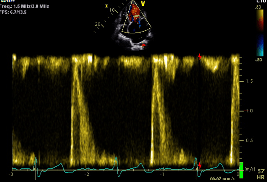
Echocardiogram
Echocardiogram is a test that looks at the heart and the surrounding blood vessels. it is one of the most important and routine investigation that is done in cardiology. Its a type of ultrasound scan, which means the heart can be seen using a small probe from the surface of your skin. The test is usually carried out by highly trained cardiac physiologists.
When is Echocardiogram used?
An echocardiogram can help with diagnosis and monitor of certain heart conditions. The test is designed to evaluate the pumping function of your heart as well as the function of its valves.
Your cardiologist may request an echocardiogram to confirm a diagnosis or as part of follow-up.
What does it involve?
For trans-thoracic echocardiogram, you will be asked to remove any clothing covering the upper half before lying down on a bed.
The physiologist will then place several small sticky sensors, known as electrodes, on the surface of your skin. These are used to monitor your heart rate and rhythm during the study.
A lubricating gel will then be applied on to your chest using a small probe. The physiologist will ask you to turn to your left hand side and may ask you to move on to your back again.
You will not hear you heart beat on the machine, however you may hear a swishing sound during the scan. This is normal and represents blood flow through your heart.
The whole test will usually take between 20-40 minutes and you will be able to go home immediately after.
Getting your results:
The results of your scan will be sent to your cardiologist who would be able to discuss them with you at your next appointment.
Are there any risks?
A standard echocardiogram is a simple and safe procedure. There are no side effects from the scan however you may have skin reaction to the electrodes which will be minimal and does not cause you any harm.


